Jazmin Camchong tests new therapies to prevent addiction relapse
By Deirdre Manion-Fischer
Relapse rates for addiction are similar to those for other chronic illnesses. At 40% - 60%, they remain high. University researchers have mapped connections in the brain that can help predict whether patients in alcohol addiction treatment programs are likely to relapse. Now, they’re working to see if they can prevent those relapses from happening.
Jazmin Camchong, an assistant professor in the Department of Psychiatry and a member of MnDRIVE and the Medical Discovery Team on Addiction, has used brain imaging to compare patterns of brain activity between healthy people and those with conditions like schizophrenia and addiction.
To map the brain, Camchong and her colleague Kelvin Lim, use functional MRI (fMRI), a technique that measures blood flow in the brain, which indicates brain activity.
For a baseline measurement, Camchong and her research team image the brain at rest. She compares this idea to taking a car in for a tune-up. “When you take your car to the mechanic, they listen to the engine when it’s just idling, and to check if all the parts are working together properly, to detect if something’s wrong,” she says, “This is what we’re doing, we’re checking whether the brain connections are there during rest.”
Camchong found that people who stayed abstinent longer demonstrate greater coordination in brain activity between the prefrontal cortex (decision-making) and the nucleus accumbens (reward center). Conversely, people with less coordination between these areas were more likely to relapse.
In an ongoing study with patients in an alcohol addiction treatment program, Camchong is working to determine whether coordination between these brain regions can be modulated to help prevent addiction relapse. Patients in the study perform a mental task that requires cognitive flexibility, basically the ability to adapt to changes.
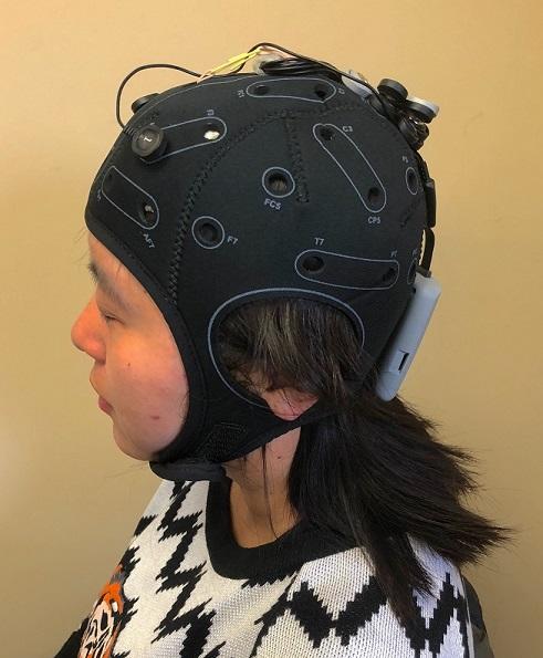
While participants perform the task, electrodes placed on the scalp deliver a mild electric current to their prefrontal cortex with a non-invasive brain stimulation technique called transcranial direct current stimulation (tDCS). The brain stimulation prepares the neurons in the prefrontal cortex to fire more readily.
Early findings indicate that participants who received brain stimulation showed higher synchrony between the prefrontal cortex and the nucleus accumbens when compared to those who did not receive brain stimulation.
The team follows up on the patients enrolled in the study several months after the tDCS sessions. Camchong found that those who received brain stimulation resumed drinking later (110 days later) than those who did not receive brain stimulation (19 days later). These are promising preliminary results for Camchong’s ongoing study.
Camchong plans to continue with this research to determine the best parameters for these types of studies, such as dose, or type of task.
The National Institute on Alcohol Abuse and Alcoholism (NIAAA) has awarded Camchong a grant to conduct a larger clinical trial. If they continue to see positive results, further research would need to verify this effect in more large clinical trials for this to become widely accepted and used in clinical settings.

Psychology undergraduate research staff member Mai Thao demonstrates wearing the tDCS device